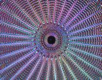
A live image of the of Diatom Arachnoidiscus under 40x magnification.

 A live image of the of Diatom Arachnoidiscus under 40x magnification.
The picture shows the diatom's silicified cell wall, which forms a pillbox-like shell (frustule) composed of overlapping halves that contain intricate and delicate markings. The picture was obtained with new video enhanced polychromatic (VEP) polscope after background subtraction. The VEP polscope shows the orientation-independent birefringence image without requiring any digital computation. An eye or camera can directly see the colored polarization image in real time through the ocular with brightness corresponding to retardance and color corresponding to the slow axis azimuth.
Image courtesy of Dr. Michael Shribak, Marine Biological Laboratory, Woods Hole, MA.
